A. Sampling
Collect the samples at the following quantities for ensuring
meaningful representation of the whole lot of feed / feedstuff
Min. sample size
Small particle type (milk, vegetable oils) 500 g
Intermediate particle type (ground meals, flours,
compounded feed)
3 kg
Small grains (wheat, rice, sorghum, ragi, barley etc.) 5 kg
Intermediate grains (maize, cotton seed / cake) 10 kg
Large grains (groundnuts / cake) 20 kg
Collect at least 100 subsamples from the whole lot. For eg. from a
truck of 100 bags of maize, collect 100 g maize from each bag to
obtain a total sample size of 10 kg
Get about 50 - 100 g subsample from the whole sample employing
either coning and quartering method (in a series of steps) or using
sample divider
The subsample thus collected can be directly subjected for analysis
B. Outline of Mycotoxin analysis
Sampling
Toxin extraction
(using organic solvents)
Clean-up
(To remove fat, impurities etc.)
Work up
Identification & Quantification
(TLC, HPLC, ELISA etc.)
C. Different methods of Mycotoxin analysis
C. 1. Thin layer chromatography (TLC)
It is the cheapest and most commonly used method. It makes use of
heterogenous equilibrium established during the flow of a solvent
(mobile phase) through a fixed phase (stationary phase) to separate ≥
2 components from materials carried by solvent (differential
migration).
Spotting the extract
Place between 5 - 20 μl of sample extract / standard as a small circular
spot (< 5 mm), 1 - 2 cm from the end of the TLC plate. Micropipette /
microcaps may be used for the purpose. Leave at least 1 cm gap
between two adjacent spots.
Developing the plate
Place about 50 - 100 ml of mobile phase (solvent) in a tank and keep
the plate at a slight angle with the spots little above the upper level of
the solvent. Due to capillary action, solvent moves upward on the
plate. Allow the solvent to travel at least about 8-10 cms.
Detection
Air dry the developed plate and view in a UV cabinet under either
longwave (365 nm) or short wave (254 nm) range to identify the
fluorescing mycotoxins. In case of mycotoxins which do not
fluoresce, spray the plate with suitable reagent to develop
fluorescence.
Resolving front value (Rf)
Each mycotoxin has its characteristic color of fluorescence under UV
light and a constant Rf value in a particular developing solvent
(Table 3). Rf value is computed using the formula,
Distance travelled by sample spot from the origin
Rf =
Distance travelled by solvent front from the origin
Confirmation
The presence of mycotoxin can be confirmed either by spraying the
plate with suitable reagents (like 50 % aqueous H2SO4, Triflouro
Acetic Acid etc.) or placing an internal standard right over the top of
the sample spot (superimposing).
Detection by Scanner
The fluorescence intensity of sample and standard spots can be
measured by using TLC Scanner / fluorodensitometer to avoid
possible human errors in comparison.
Table 3. TLC characteristics of mycotoxins
Toxin Rf * Color Color (UV) after
(UV) spray * *
Aflatoxin B1 0.31 Blue Pink
Aflatoxin B2 0.26 Blue Pink
Aflatoxin G1 0.23 Green Blue
Aflatoxin G2 0.17 Green Blue
Ochratoxin A 0.55 Green Blue
T-2 toxin 0.36 Yellow Blue
Zearalenone 0.78 Blue Yellow
DAS 0.33 Yellow Variable
Sterigmatocystin 0.85 Red-brown Yellow
*TEF : Toluene : ethyl acetate : formic acid ( 6:3:1 )
* *P - anisaldehyde
C. 2. Spectrophotometry
This is an extension of TLC method. The sample spots on the
developed TLC plate are scraped out alongwith the sorbent (silica gel)
and extracted with methanol for 3 minutes. The extract is filtered and
the absorbance of the filtrate is measured in a spectrophotometer
at 363 nm.
Reference :
Nabney and Nesbitt. 1965. Analyst 90 : 155-160.
C. 3. High Performance Thin Layer Chromatography (HPTLC)
This is an improvised version of TLC, where sampleapplication and detection of fluorescence intensity are fully automated
and carried out by using automated sample applicator (like Linomat
IV of Camag, Switzerland) and densitometer, respectively.
Mycotoxin levels less than 0.1 ppb can be detected by this method.
C. 4. Minicolumn method
A glass column of 20 cm length, 6 mm internal diameter withtapering end (2 mm) is packed serially from the bottom with glass
wool, calcium or sodium sulphate (8-10 mm), florisil (8-10 mm),
silica gel (18-20 mm), neutral aluminia (8-10 mm), calcium or
sodium sulphate (8-10 mm) and a cap of glass wool.
2 ml of final chloroform extract (in case of aflatoxin) is placed
in the column and eluted with chloroform : acetone (9 : 1). Aflatoxin,
if present is trapped as a band above florisil layer which can be
viewed under long wave UV light as a blue fluorescent band. This
method can be used as a qualitative test for rapid identification of
mycotoxin.
C. 5. Immuno assays
These assays are developed on the basic principle ofAntigen - Antibody reaction. Antibodies are highly specific to the
Mycotoxin - Protein conjugate (Hapten) used. Hence the results will
be highly specific.
Commonly employed immuno assays
Radio immuno assay (RIA)
Standard mycotoxin, labelled onto a radioactive compound like
Tritium is used. Mycotoxin levels as low as 2-5 ppb can be
detected. The disadvantages of this method include high cost,
difficulty in labelling, radio active waste disposal problem and
risk of handling.
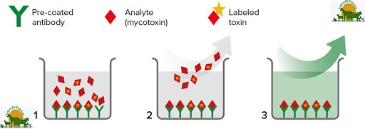
Enzyme linked immuno sorbent assay (ELISA)
It has received great attention in recent times and has been the
most popular and widely practiced immuno assay method.
ELISA is rapid, more sensitive, highly specific and simple to
operate. It does not require any extensive extraction or cleanup.
Commercial ELISA kits
Various companies have been marketing commercial kits
which basically work on ELISA principle. These have gained
wider acceptance as considerable amount of time is saved on
antibody production. Sample is extracted with methanol : water
(60 : 40) or acetonitrile : water (50 :50) and the extract is
directly subjected to analysis.
Elisa tests are good for quick identification of
mycotoxins in feed samples, various tests are developed based
on Antigen - Antibody principle. Some companies which
produce ELISA kits are :
1. Neogen Corp,
620, Lesher place,
Lansing, Michigan 48912, U.S.A.
2. Vicam,
313, Pleasant St.,
Watertown, Massachusetts - 02172, U.S.A.
C. 6. High performance liquid Chromatography (HPLC)
It is highly sensitive and can detect upto 5 x 10⁻⁶ ppb level ofmycotoxin. Stainless steel columns (< 18) of 15 cm length and 4 mm
internal diameter, packed with silica gel (particle size - 5 microns) are
used. Sample is first extracted with suitable solvent (generally 60 %
aqeous methanol) and the extract is cleaned - up.
This purified extract (20 μl) is injected into the column and the
eluent (generally a mixture of methanol, water and acetonitrile) is
passed at a flow rate of 0.75 ml / min and at a pressure of 3000 psi.
The eluted toxins coming out of the column are detected and
quantified by fluorimeter.
The columns may be either normal phase (polar stationary
phase) or reverse phase (polar mobile phase) type. The latter type is
most commonly used.
C. 7. Bio - assays
Mostly are useful as confirmatory tests. Toxin extract isinjected as a single dose into stomach (day-old duckling bioassay,
guinea pig bioassay), fertile eggs (chick embryo bioassay) or into skin
of rabbits (skin bioassay). Presence of toxin is confirmed by noticing
pathological changes or mortality.
Safety precautions in mycotoxin analysis
Carryout the mycotoxin analysis in a separate work area in the
laboratory
Cover the bench top with non absorbent material
Solvents used are highly inflammable. So avoid using electric stoves,
bunsen burners etc.
Do not stock the solvents in larger quantities
Wear protective clothing, gloves and mask to minimise the risk of
inhalation / contact with hazardous mycotoxins
Some of the solvents (like benzene, chloroform) are toxic. Avoid
direct skin contact with them
Any spillage should be immediately mopped-up with cotton. Such
cotton should be incinerated
After completing the work, decontaminate the area with 4 % sodium
hypochlorite solution
Decontaminate the glassware by soaking for atleast 2 hours in 1 %
sodium hypochlorite solution
Spray the TLC plate with reagent only in a fume cup-board / spray
cabinet
At the UV cabinet, always view the TLC plate only through the UV
filter
Avoid eating, drinking and smoking in the laboratory
Keep the lab well ventilated using exhaust fans
