Pages

Tuesday 28 January 2020

Definitions

Nutrition:
            It is the science of series of process by which food or feed is taken in and absorbed into the body of an organism which serves for purpose of growth, work , maintenance and repair of the vital process.

Nutrient:
            It may be any feed constituent that aids in the support of life.

Concentrate:
            The feed stuffs which are rich in TDN and low in fibre contents.

Roughages:
            The feed stuffs which are rich in crude fibre and low in total digestible nutrients.

Forage:
            Forage is a broad term. Any roughages used for livestock feed including fodder, pasture, range land grasses and straws.

Fodder:
            Fodder is a part of forage. It is only cultivated forage which is cut and offered to animals.
Hay:
            Green forage harvested during the growing season and preserved by drying for subsequent use during fodder scarcity period.

Silage:
Silage is green plant material preserved by anaerobic fermentation.

Pasture:
            A fenced area of domesticated forage, usually improved on which animal are grazed.

Range:
Large, naturally vegetated area of relatively low productivity unfenced grazed by livestock.

Total Digestible Nutrients:
            A term used to express the energy value of feed stuffs or feed mixture. It is determined by the summation of the digestible C.P+digestible C.F+ digestible E-E/time*2.25 and + digestible NFE. It express amount of heat or energy present in feed stuffs.


Non Protein Nitrogen:
            Nitrogen originating from other than an amino acid sources but may be used by bacteria in the rumen to synthesize protein NPN sources includes compounds like urea and anhydrous ammonia which are used in feed formulation for ruminants.

Microbiology:

Allergy:
            It is a hypersensitive state acquired through exposure to particular allergen exposure eliciting an altered capacity to react.

Antibody:
            It is the immunoglobulin product of B-cells and plasma cells that combines specially with the antigen that activated the cell.

Antigen:
            It is the substance that activates the immune system to produce T-cells or B- cells against that substance.

Asepsis:
            It is the freedom from infections

Antiseptic:
            These are the substances which kill or prevent the growth of microorganisms, which applied locally on living tissues. Like povidine etc.

Disinfectants:
            These are the substance which kill or prevent the growth of microorganisms, which are applied on living things. Like phenols etc.

Vaccine:
            It is a suspension of attenuated or killed microorganisms administered for the prevention, or treatment of infectious diseases.

Attenuated Virus:
            It is one whose pathogenicity has been reduced by serial animal passage or by other means.

Acquired Immunity:
            It is the state of heightened specific immunity acquired by exposure to a particular foreign antigen.

Endotoxin:
            The toxin produced after the death of bacteria is known as endotoxin.

Exotoxin:
            The toxin produced by live bacteria is known as endotoxin.

Animal Breeding And Genetics:
Acquired Character:
            This term applies to possibilities of an environmentally induced change in body becoming hereditary

Aberration:
            A change from the normal  is known as aberration.

Alleles:
            Alternative forms of genes are called alleles.

Correlation:
            Association between characteristics of individuals is known as correlation.

Covariance:
            Variation that is common between two traits. It may be result from joint hereditary or environmental influences.

Genotype:
            This is the complete genetic make up of an individual.

Phenotype:
            It is the external appearance or some other observable or measurable characteristics of an individual.

Pedigree:
            It is a record of animals from which a given individual is descended. The definition is always extended to include animals, which are collaterally related to an individual.

Progeny:
            The young or offspring of the given individuals.


Selection:
            The causing or allowing certain individuals to produce the next generation.

Sire:
            Sire is the father of an individual.

Calving Interval:
            The period between birth of two successive calves from one cow.

Dry Period:
            Period of non-lactating between two periods of lactation.

Service Period:
            The time from calving to next conception is known as service period.

Gestation Period:
            The period from mating to the parturition is known as gestation period.

Proven Sire:
            A bull with at least 10 daughters which have completed lactation records and which are born of dams with completed lactation records.

Animal Reproduction:

Abortion:
            It is the expulsion of dead fetus or recognizable size at any stage of gestation.

Agalactia:
            The absence of milk in the udder of freshly parturated dam.

Dystokia:
            Abnormal or difficult birth is called dystokia.

Eutokia:
            It is normal birth of child.


Temperature, Pulse Rate and Respiration Rate (TPR)

Animal
Normal temp (oF)
Normal Pulse rate/min
Normal Respiration/min
Buffalo
100.5
40-60
15-20
Cattle
102
60-70
15-25
Sheep
102
60-70
15-30
Goat
102.5
70-80
15-30
Horse
100
28-40
10-14
Camel
99.5
32-44
5-12
Dog
102
65-90
20-30
Cat
101.5
110-130
30-40
Poultry
107
120-140
15-30

     Formula to convert oF in oC.
oC=(oF-32)/9*5


Age of Puberty in different Animals


Animal
Age of puberty
Cattle
12-22 month
Buffalo
36 months
Horse
12-24 month
Sheep
8-12 month
Goat
6-10 month
Camel
12-24 month
Dog
6-12 month
Cat
7-15 month
Poultry
20-22 week


Estrous Cycle Length in different animals


Species
Classification
Estrous cycle
Cow/Buffalo
Polyestrus
21 days
Mare
Seasonal polyestrus (long day)
21 days
Ewe/Doe
Seasonal polyestrus (short day)
17 days
Queen
Polyestrus
17 days
Bitch
Monoestrus
6 months


Gestation length in different animals

Species
Gestation periods in months
Gestation period in days
Cattle
09±09
270±10 days
Buffalo
10±10
305±10 days
Ewe/Doe
05±05
150±5 days
Mare
11±10
330±10 days
Camel
12±12
365±12 days
Bitch
2±10
60±10 days
Queen
02±10
60±10 days

Average age of different animals

Species
age in years
Cow
15-20 years
Sheep
7-9 years
Goat
8-10 years
Horse
30-50 years
Dog
10-15 years
Cat
09-10 years
Deer
15 years
Camel
30-35 years

Incubation  Period of different Birds

Species
Incubation period
average incubation period
Chicken
20-22 days
21 days
Duck
26-28 days
27 days
Goose
30-33 days
32 days
Turkey
26-28 days
27 days
Parrot
17-31 days
24 days
Pigeon
16-18 days
17 days
Quill
21-28 days
25 days

Rapid TLC Method of Multi-Mycotoxin Analysis (Modified Tapia Method)


7. Rapid TLC Method of Multi-Mycotoxin Analysis (Modified Tapia Method)
Reagents :
i) 4 % KCl (4 g KCl in 100 ml di. water)
ii) 5 N HCl (405.9 ml Conc. HCl in 1 l di. water)
iii) Na2So4 ( anhydrous )
iv) 20 % KOH (20 g KOH in 100 ml di. water)
v) 20 % H2SO4 in ethanol (20 ml Conc. H2SO4 + 80 ml ethanol)

Solvents :

  1. Acetonitrile
  2. Hexane
  3. Chloroform
  4. Chloroform : Acetone : Water (88 : 12 : 1)
  5. Toluene : Ethyl acetate : Formic acid (5 : 4 : 1)


Standards : 

  • Aflatoxin B11 µg/ml in Acetonitrile: Benzene (2 : 98)
  • Ochratoxin A 2 µg/ml in Acetonitrile: Benzene (2: 98)
  • Zearalenone 50 µg / ml in Benzene
  • T-2 toxin 50 µg / ml in Ethyl acetate

         Sterigmatocystin         Citrinin            Oosporein

Procedure :

  • Take 25 g sample, add 85 ml acetonitrile, 15 ml 4 % KCl and 2 ml 5 N HCl and blend at high speed for 3 minutes
  • Filter through Whatman no. 1 filter paper
  • Transfer 50 ml filtrate into a 250 ml separating funnel
  • Add 50 ml water, followed by 50 ml hexane and shake well
  • Collect the lower layer, add 50 ml hexane again and repeat the above step
  • Collect the lower layer into another separating funnel and extract with two 10 ml portions of chloroform
  • Drain the chloroform layer through anhydrous Sodium sulphate and evaporate in oven at 50 oC
  • Dissolve the residue in 0.2 ml chloroform and spot on TLC plate along with the standards
  • Develop plate in chloroform : acetone : water (88 : 12 : 1) in one direction and toluene : ethyl acetate : formic acid (5 : 4 : 1) in the second direction
  • Spray zone of spots, corresponding to sterigmatocystin with 20 % aqueous KOH
  • Spray zone of spots, corresponding to T-2 toxin with 20 % H2So4 in ethanol and heat at 110C
  • View the spots, identify and quantify the toxins as done with individual toxins                                                                                                                                                                                Effective weight of the sample = 25 x 50 = 12.255 g

ANALYSIS OF CITRININ


ANALYSIS OF CITRININ


Reagents:
1. 4% KCL (4 gms KCL in 100 ml distilled water)
2. 20% H2SO4 (20 ml concentrated H2SO4 + 5% NaHC03( 5 gms of NaHC03 in 100 ml DW)
3. 6 N HCl (185.4 ml of HCl in 1 lt distilled water)
4. 10% Oxalic acid in methanol (W/V) (10 gm oxalic acid in 100 ml methanol)

Solvents: 
1. Acetonitrile
2. Iso octane
3. Chloroform
4. Chloroform – methanol – hexane (64:1:35)

Standards:
1. Citrinin : 10 g/ml in methanol

Procedure:

  • Take 25 gms of sample, add 180 ml acetonitile, 20 ml 4% KCl, 2 ml 20% H2SO4 and shake for 15 minutes
  • Filter and collect 100 ml of the filtrate
  • Transfer filtrate to separating funnel and add 50ml iso octane. Shake for 1 minute.
  • Collect the lower layer, add 50 ml iso octane and repeat the above step.
  • Collect the lower layer into another separating funnel, add 25 ml water and extract with 50 ml chloroform
  • Drain the chloroform layer into a separating funnel
  • Extract again with two 10 ml portion of chloroform and combine all the three extracts
  • Add 25 ml of 5% NaHC03 to the extract. Shake for 1 minute and drain off the lower portion.
  • Re-extract with two 25 ml 5% NaHC03 portion and repeat the above step.
  • Combine the extracted portion in a 600 ml beaker and acidify it with 6N HCl to pH 1-2, transfer it to separating funnel
  • Rinse the beaker with 50 ml CHCl3, and transfer it to seperating funne, swirl for 30 seconds.
  • Drain the lower portion, repeat above step with another 50 ml portion of CHCl3
  • Collect the CHCl3 extracts and evaporate to near dryness
  • Add 5 ml CHCl3 to the dried extract and filter through fluorocarbon filter into a 25 ml beaker
  • Rinse the beaker twice with small amounts of CHCl3, filter and evaporate
  • Dip the TLC plate in 10% oxalic acid solution for 2 min and air dry overnight
  • Spot the sample and standard in 5-20 l range
  • Develop plate in chloroform-methanol-hexane (64:1:35) for 45 minutes. Air dry and observe under long wave UV light for comparing the intensitites
  • Calculate the citrinin content of sample using the following formula

Citrinin, ug/kg = S x Y x V X x W

Where,
           S = ul standard equal to unknown
           Y = Concentration of standard (ug/ml)
           V = Dilution of sample extract (ul)
           X = ul of sample spotted
           W= gms of sample represented by final extract

 = 25 x 100 = 12.376 g 202

Rapid TLC method of Aflatoxin analysis


3. Rapid TLC method of Aflatoxin analysis
(Modified Romer's method)

Reagents :
 i) 0.2 M NaOH (dissolve 8 g NaOH in water and
make up volume to 1 lit)
ii) 0.41 M Ferric Chloride (dissolve 66.5 g anhydrous
FeCl3 in water and make up volume to 1 lit)
iii) 0.03 % H2SO4 (0.3 ml conc. H2SO4 + 999.7 ml
water)
iv) Potassium wash solution (dissolve 1.12 g KOH
and 10 g KCl in water and make up volume to 1
lit)

Solvents :
 i) Acetone
ii) Chloroform
iii) Developing solvent
Chloroform : Acetone : Water (88 : 12 : 1)
Standard : Aflatoxin B1 - 1 μg/ml in Benzene : Acetonitrile (98:2)

Procedure :
 Take 25 g sample in a conical flask, add 100 ml distilled water and
blend for 2 minutes
 Add 150 ml acetone and blend again for 2 minutes
 Filter through Whatman no. 1 filter paper and transfer 75 ml of filtrate
to a conical flask containing 3 g cupric carbonate
 Prepare ferric gel by adding 85 ml of 0.2 M NaOH to 15 ml of 0.41 M
FeCl3. Add this mixture to the flask containing extract and cupric
carbonate
 Mix the contents slowly by swirling movements
 Filter through Whatman no. 1 filter paper
 Take 100 ml of filtrate in a 250 ml separating funnel
 Add 100 ml of 0.03 % H2SO4 and 10 ml of chloroform. Mix the
contents slowly
 Collect the chloroform layer into a 100 ml beaker
 Add again 10 ml of chloroform to the separating funnel and repeat the
above step. Combine both the chloroform extracts
 Take 100 ml potassium wash solution in a separate separating funnel
 Add the chloroform extract to the second separating funnel and mix it
slowly
 Collect the chloroform layer through anhydrous sodium sulfate bed
drop by drop to remove moisture
 Dry the chloroform extract in an oven at 50°C
 Dissolve the dried residue in 0.2 ml chloroform and spot on TLC plate
along with the standard
 Compare the flourescence intensities of the sample and standard spots
and identify the ones matching with each other
 Calculate the aflatoxin content in the following way

          Aflatoxin content (ppb) =                       S x C x D   x 1000
T x W
Where, S = Standard which compares with the sample in
fluorescent intensity
C = Concentration of standard (1 μg / ml)
D = Dilution factor in ml
T = Sample which compares with standard in fluorescent
intensity

                              W = Effective weight [25 x  75 x 100     = 4.286 g]
250 x 175


4. Analysis of Ochratoxin
(By thin layer chromatography)

Reagents : 
Sodium bicarbonate and diatomaceous earth mixture :
Add 25 ml of 5 % aqueous NaHCO3 to 50 g
diatomaceous earth (Celite 545), mix well and store in
tightly closed container

Solvents :
 i) Chloroform
ii) Hexane
iii) Acetic acid : benzene ( 2 : 98 )
iv) Acetic acid : benzene (1 : 99 )

Standard : 
Ochratoxin A 2 μg / ml in acetic acid : benzene (1 : 99)

Apparatus :
 i) Wrist action / horizontal shaker
ii) Hot water (steam) bath
iii) TLC plates (precoated silica gel plates or
equivalent)
iv) Developing tank / chamber
v) UV viewing cabinet

Procedure :
 Take 25 g of sample in a 250 ml glass stoppered conical flask, add
12.5 ml water and mix
 Add 125 ml chloroform and shake for 1 hour
 Filter through Whatman No.1 filter paper and collect the filtrate
 Plug the bottom of a glass column (2 cm x 30 cm) with glass wool,
put 6 g of NaHCO3 - Celite mixture and tamp firmly with a glass rod
 Add 50 ml of chloroform extract to the column and elute until
meniscus reaches top of the NaHCO3 - Celite column
 Wash the column with 70 ml hexane followed by 70 ml chloroform
and discard washings
 Elute Ochratoxin with 100 ml acetic acid : benzene ( 2 : 98)
 Collect the eluate and evaporate on steam bath
 Dissolve the residue in 5 - 10 ml chloroform, transfer to a small vial
( 10 ml capacity) and evaporate on steam bath
 Dissolve the residue in 0.5 ml acetic acid : benzene (1 : 99) by
vigorous shaking
 Spot on TLC plate along with the standard (5, 10, 15 and 20 μ1 or in
any other suitable range)
 Develop the plate using toluene : ethyl acetate : formic acid ( 5 : 4 :
1) in an unequilibrated chamber
 Air dry the plate, view under long wave UV light (365 nm) and
compare the intensity of greenish blue fluorescent spots of the sample
with that of standard spots and identify the spot, matching each other
 Calculate the Ochratoxin A content using the formula
                                   Ochratoxin A μg / kg = S x Y x V
Z x W
Where, S = Volume in μl of ochratoxin A standard spot
comparable to Z μl of sample spot
Z = Volume in μl of sample spot comparable to S μl of
ochratoxin A standard
Y = Concentration of ochratoxin Astandard (2 μg / ml)
V = Volume (μl) of the dissolved residue before spotting
W= Effective weight of the sample
25 x 50
150

Confirmation

Expose the developed plate to NH3 fumes. Greenish blue fluorescence
of Ochratoxin will turn to bright blue.

Reference

AOAC. 1995. Official methods of analysis. 16th ed. Assoc. Off. Anal.
Chem., Washington, D.C.

5. Analysis of T-2 toxin
(By thin layer chromatography)

Reagents : 
i) 30 % ammonium sulphate (dissolve 30 g (NH4)2
SO4 in water and make up volume to 100 ml)
ii) Celite 545
iii) Potassium wash solution (dissolve 1.12 g KOH
and 10 g KCl in water and make up volume to 1 lit)
iv) Sodium sulphate
v) Silica gel
vi) Methanol : H2SO4 ( 1 : 1 v/v )

Solvents : 
i) Methanol : water (1 : 1 v/v)
ii) Chloroform
iii) Diethyl ether
iv) Hexane
v) Benzene
vi) Acetone : Benzene (5 : 95 v/v)
vii) Developing solvent mixture - Toluene : ethyl
acetate formic acid (6 : 3: 1 v/v)

Standard :
T-2 toxin 50 μg / ml in Benzene or diethyl ether

Apparatus : 
i) Wrist action / horizontal shaker
ii) TLC plates (precoated silica gel plates
or equivalent)
iii) Developing tank / chamber
iv) UV viewing cabinet

Procedure :
 Take 50 g of sample in a glass stoppered conical flask
 Add 250 ml of methanol : water (1 : 1) and shake for 1 hour
 Filter using whatman No.1 filter paper and collect 60 ml of extract
into a beaker
 Add 240 ml 30 % (NH4)2 SO4 and stir vigorously for 1 minute
 Add 20 g of celite and stir for 1 minute
 Filter and collect 200 ml of filtrate
 Transfer filtrate to a separating funnel
 Add 10 ml of chloroform and shake vigorously for 1 minute
 Allow the layers to separate and collect the bottom layer into another
separating funnel
 Repeat the extraction with another 10 ml of chloroform
 Combine both the extracts and add 100 ml of potassium wash solution
 Swirl gently for 30 seconds and let layers separate
 Drain the lower chloroform layer through a bed of Sodium sulphate
(in a funnel) to dry and collect 10 ml of clear filtrate
 Column Preparation : Plug the bottom of a glass column ( 2 cm x
30 cm ) with glass wool and add 5 g anhydrous sodium sulphate. Fill
the column to half level with chloroform and add 10 g silica gel.
Wash sides of column with chloroform and stir to eliminate air
bubbles. Drain off chloroform leaving about 7 cm above the upper
level of silica gel. Add 15 g anhydrous sodium sulphate without
disturbing the silica gel. Drain off chloroform to the upper level of
sodium sulphate
 Wash the column serially with 50 ml of diethyl ether and 10 ml of
chloroform and discard the washings
 Mix 10 ml of sample extract with 30 ml of hexane and add to the
column and slowly drain until solvent is about 1 cm above Sodium
sulphate
 Add in succession 30 ml benzene and 40 ml acetone : benzene (5: 95)
and discard both the washings
 Elute T-2 with diethyl ether until 30 ml of eluate is collected and
evaporate the eluate
 Dissolve the residue in 0.5-1.0 ml diethyl ether. Spot on TLC along
with the standard (5-20 μ1 or any other suitable range) and develop
the plate in toluene : ethyl acetate : formic acid (6 : 3 : 1)
 Air dry the plate and spray with methanol : H2SO4 (1 : 1)
 Dry at 110°C for 10 minutes and observe blue fluorescence under
long wave UV light (365 nm)
 Compare the intensities of the blue fluorescent spots of the sample
with those of standard and identify the ones matching each other
 Calculate the T-2 content of sample using the following formula
T - 2 μg / kg =    S x Y x V
                            Z x W
Where, S = Volume in μl of T - 2 standard spot comparable to
Z μl of sample spot
Z = Volume in μl of sample spot comparable to S μl of
T - 2 standard
Y = Concentration of T - 2 standard (50 μg / ml)
V = Volume (μl) of the dissolved residue before spotting
W= Effective weight of the sample
       50 x 60 x 200     x 10
                                                                  250   300    20

References
Romer, T.R., Boling, T.M. and Mc Donald, J.L. 1978. Gas liquid
chromatographic determination of T-2 toxin and diacetoxyscirpenol in
corn and mixed feeds. JAOAC. 61 : 801 - 807.
Rukmini, C. and Bhat, R.V. 1978. Occurrence of T-2 toxin in Fusarium
infested sorghum from India. J. Agric. Food Chem. 26 : 647-649.

6. Analysis of Zearalenone
(By thin layer chromatography)
Reagents :
 i) Aluminium chloride solution (dissolve 20g
AlCl3 6H2O in 100 ml methanol)
ii) Celite 545

Solvents : 
i) Chloroform : water mixture ( 10 : 1 v/v)
ii) Hexane
iii) Chloroform
iv) Diethyl ether
v) Benzene
vi) Acetone : benzene ( 5 : 95 v/v)
vii) Acetonitrile
viii) Developing solvent -
Methanol : Chloroform (5 : 95 v/v) or
Acetic acid : Benzene (5 : 95 v/v)

Standard :
Zearalenone 50 μg / ml in Benzene

Apparatus :
i) Wrist action / horizontal shaker
ii) Hot water (steam) bath
iii) TLC plates (precoated silica gel plates or
equivalent)
iv) Developing tank / chamber
v) UV viewing cabinet

Procedure :
 Take 50 g of sample in a glass stoppered conical flask and add 300 ml
of chloroform : water (10 : 1) and 25 g of celite
 Shake for 1 hour and filter using Whatman No. 1 filter paper
 Column Preparation : Plug the bottom of a glass column
(2 cm x 30 cm) with glass wool and add 5 g anhydrous sodium
sulphate. Fill the column about half full with chloroform and add 10 g
silica gel. Wash sides of column with chloroform and stir to
eliminate air bubbles. Drain off chloroform leaving about 7 cm above
the upper level of silica gel. Add 15 g anhydrous sodium sulphate
without disturbing the silica gel. Drain off chloroform to the upper
level of sodium sulphate
 Transfer 50 ml of sample extract together with 150 ml hexane into the
silica gel column
 Drain until the solvent reaches top of the column and discard the
washings
 Wash the column serially with 150 ml of diethyl ether and 150 ml of
benzene and discard both the washings
 Elute Zearalenone with 250 ml of acetone : benzene ( 5 : 95 )
 Add few silica chips to the eluate and evaporate on steam bath,
preferably under gentle stream of N2
 Dissolve residue in 10 ml of hexane and transfer quantitatively to a
separating funnel
 Repeat the above step for 3 times using 10 ml of hexane each time
 Finally rinse the residue with 10 ml of acetonitrile and add to the
hexane washes present in the separating funnel. Shake well and let
phases separate
 Collect the lower acetonitrile phase into a 100 ml beaker
 Add another 5 ml acetonitrile to the hexane washes present in the
separating funnel and repeat the above step. Combine both the
acetonitrile fractions.
 Evaporate the combined acetonitrile fractions on steam bath ( under
stream of N2)
 Transfer the residue to a small vial (about 10 ml capacity) using about
5 - 10 ml of chloroform and evaporate as in the previous step
 Add about 0.5 ml benzene to the residue and shake vigorously
 Spot on TLC plate (5, 10, 15, 20 μl or other suitable volumes) along
with the standard and develop the plate in methanol : chloroform
(5 : 95) or acetic acid : benzene (5 : 95)
 Air dry the plate and spray the spots with aluminium chloride
solution, heat at 130° C for 5 min and examine under longwave UV
light (365 nm)
 Compare the intensities of the blue fluorescent spots of sample with
those of the standard and identify the ones, matching with each other.
Calculate the Zearalenone content of the sample in the following way
Zearalenone μg / kg = S x Y x V
                                     Z x W
Where, S = Volume in μl of Zearalenone standard spot
comparable to Z μl of sample spot
Z = Volume in μl of sample spot comparable to S μl of
Zearalenone standard
Y = Concentration of Zearalenone standard (1 μg / ml)
V = Volume (μl) of the dissolved residue before spotting
W= Effective weight of the sample
50x   50
         300
References :
AOAC, 1995. Official methods of analysis. 16th ed. Assoc. Off. Anal.
Chem. Washington, D.C.

Mycotoxin Analysis


A. Sampling

 Collect the samples at the following quantities for ensuring
meaningful representation of the whole lot of feed / feedstuff
Min. sample size
Small particle type (milk, vegetable oils) 500 g
Intermediate particle type (ground meals, flours,
compounded feed)
3 kg
Small grains (wheat, rice, sorghum, ragi, barley etc.) 5 kg
Intermediate grains (maize, cotton seed / cake) 10 kg
Large grains (groundnuts / cake) 20 kg
 Collect at least 100 subsamples from the whole lot. For eg. from a
truck of 100 bags of maize, collect 100 g maize from each bag to
obtain a total sample size of 10 kg
 Get about 50 - 100 g subsample from the whole sample employing
either coning and quartering method (in a series of steps) or using
sample divider
 The subsample thus collected can be directly subjected for analysis

B. Outline of Mycotoxin analysis
Sampling

Toxin extraction
(using organic solvents)
Clean-up
(To remove fat, impurities etc.)
Work up
Identification & Quantification
(TLC, HPLC, ELISA etc.)

C. Different methods of Mycotoxin analysis

C. 1. Thin layer chromatography (TLC)


 Principle

It is the cheapest and most commonly used method. It makes use of
heterogenous equilibrium established during the flow of a solvent
(mobile phase) through a fixed phase (stationary phase) to separate ≥
2 components from materials carried by solvent (differential
migration).

 Spotting the extract

Place between 5 - 20 μl of sample extract / standard as a small circular
spot (< 5 mm), 1 - 2 cm from the end of the TLC plate. Micropipette /
microcaps may be used for the purpose. Leave at least 1 cm gap
between two adjacent spots.

 Developing the plate

Place about 50 - 100 ml of mobile phase (solvent) in a tank and keep
the plate at a slight angle with the spots little above the upper level of
the solvent. Due to capillary action, solvent moves upward on the
plate. Allow the solvent to travel at least about 8-10 cms.

 Detection

Air dry the developed plate and view in a UV cabinet under either
longwave (365 nm) or short wave (254 nm) range to identify the
fluorescing mycotoxins. In case of mycotoxins which do not
fluoresce, spray the plate with suitable reagent to develop
fluorescence.

 Resolving front value (Rf)

Each mycotoxin has its characteristic color of fluorescence under UV
light and a constant Rf value in a particular developing solvent
(Table 3). Rf value is computed using the formula,
Distance travelled by sample spot from the origin
Rf =
Distance travelled by solvent front from the origin

 Confirmation

The presence of mycotoxin can be confirmed either by spraying the
plate with suitable reagents (like 50 % aqueous H2SO4, Triflouro
Acetic Acid etc.) or placing an internal standard right over the top of
the sample spot (superimposing).

 Detection by Scanner

The fluorescence intensity of sample and standard spots can be
measured by using TLC Scanner / fluorodensitometer to avoid
possible human errors in comparison.

Table 3. TLC characteristics of mycotoxins
Toxin Rf * Color Color (UV) after
(UV) spray * *
Aflatoxin B1 0.31 Blue Pink
Aflatoxin B2 0.26 Blue Pink
Aflatoxin G1 0.23 Green Blue
Aflatoxin G2 0.17 Green Blue
Ochratoxin A 0.55 Green Blue
T-2 toxin 0.36 Yellow Blue
Zearalenone 0.78 Blue Yellow
DAS 0.33 Yellow Variable
Sterigmatocystin 0.85 Red-brown Yellow

*TEF : Toluene : ethyl acetate : formic acid ( 6:3:1 )
* *P - anisaldehyde

C. 2. Spectrophotometry

This is an extension of TLC method. The sample spots on the
developed TLC plate are scraped out alongwith the sorbent (silica gel)
and extracted with methanol for 3 minutes. The extract is filtered and
the absorbance of the filtrate is measured in a spectrophotometer
at 363 nm.

Reference :

Nabney and Nesbitt. 1965. Analyst 90 : 155-160.

C. 3. High Performance Thin Layer Chromatography (HPTLC)
This is an improvised version of TLC, where sample
application and detection of fluorescence intensity are fully automated
and carried out by using automated sample applicator (like Linomat
IV of Camag, Switzerland) and densitometer, respectively.
Mycotoxin levels less than 0.1 ppb can be detected by this method.

C. 4. Minicolumn method
A glass column of 20 cm length, 6 mm internal diameter with
tapering end (2 mm) is packed serially from the bottom with glass
wool, calcium or sodium sulphate (8-10 mm), florisil (8-10 mm),
silica gel (18-20 mm), neutral aluminia (8-10 mm), calcium or
sodium sulphate (8-10 mm) and a cap of glass wool.
2 ml of final chloroform extract (in case of aflatoxin) is placed
in the column and eluted with chloroform : acetone (9 : 1). Aflatoxin,
if present is trapped as a band above florisil layer which can be
viewed under long wave UV light as a blue fluorescent band. This
method can be used as a qualitative test for rapid identification of
mycotoxin.
C. 5. Immuno assays
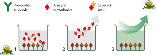
These assays are developed on the basic principle of
Antigen - Antibody reaction. Antibodies are highly specific to the
Mycotoxin - Protein conjugate (Hapten) used. Hence the results will
be highly specific.

Commonly employed immuno assays

 Radio immuno assay (RIA)
Standard mycotoxin, labelled onto a radioactive compound like
Tritium is used. Mycotoxin levels as low as 2-5 ppb can be
detected. The disadvantages of this method include high cost,
difficulty in labelling, radio active waste disposal problem and
risk of handling.

 Enzyme linked immuno sorbent assay (ELISA)
It has received great attention in recent times and has been the
most popular and widely practiced immuno assay method.
ELISA is rapid, more sensitive, highly specific and simple to
operate. It does not require any extensive extraction or cleanup.

Commercial ELISA kits
Various companies have been marketing commercial kits
which basically work on ELISA principle. These have gained
wider acceptance as considerable amount of time is saved on
antibody production. Sample is extracted with methanol : water
(60 : 40) or acetonitrile : water (50 :50) and the extract is
directly subjected to analysis.
Elisa tests are good for quick identification of
mycotoxins in feed samples, various tests are developed based
on Antigen - Antibody principle. Some companies which
produce ELISA kits are :
1. Neogen Corp,
620, Lesher place,
Lansing, Michigan 48912, U.S.A.
2. Vicam,
313, Pleasant St.,
Watertown, Massachusetts - 02172, U.S.A.
C. 6. High performance liquid Chromatography (HPLC)
It is highly sensitive and can detect upto 5 x 10⁻⁶ ppb level of
mycotoxin. Stainless steel columns (< 18) of 15 cm length and 4 mm
internal diameter, packed with silica gel (particle size - 5 microns) are
used. Sample is first extracted with suitable solvent (generally 60 %
aqeous methanol) and the extract is cleaned - up.
This purified extract (20 μl) is injected into the column and the
eluent (generally a mixture of methanol, water and acetonitrile) is
passed at a flow rate of 0.75 ml / min and at a pressure of 3000 psi.
The eluted toxins coming out of the column are detected and
quantified by fluorimeter.
The columns may be either normal phase (polar stationary
phase) or reverse phase (polar mobile phase) type. The latter type is
most commonly used.
C. 7. Bio - assays
Mostly are useful as confirmatory tests. Toxin extract is
injected as a single dose into stomach (day-old duckling bioassay,
guinea pig bioassay), fertile eggs (chick embryo bioassay) or into skin
of rabbits (skin bioassay). Presence of toxin is confirmed by noticing
pathological changes or mortality.
Safety precautions in mycotoxin analysis
 Carryout the mycotoxin analysis in a separate work area in the
laboratory
 Cover the bench top with non absorbent material
 Solvents used are highly inflammable. So avoid using electric stoves,
bunsen burners etc.
 Do not stock the solvents in larger quantities
 Wear protective clothing, gloves and mask to minimise the risk of
inhalation / contact with hazardous mycotoxins
 Some of the solvents (like benzene, chloroform) are toxic. Avoid
direct skin contact with them
 Any spillage should be immediately mopped-up with cotton. Such
cotton should be incinerated
 After completing the work, decontaminate the area with 4 % sodium
hypochlorite solution
 Decontaminate the glassware by soaking for atleast 2 hours in 1 %
sodium hypochlorite solution
 Spray the TLC plate with reagent only in a fume cup-board / spray
cabinet
 At the UV cabinet, always view the TLC plate only through the UV
filter
 Avoid eating, drinking and smoking in the laboratory
 Keep the lab well ventilated using exhaust fans

MYCOTOXIN QUANTIFICATION


1. Mycotoxin standards
Pure mycotoxins of known concentration are used in mycotoxin
assays for either fluorescence intensity comparison or calibration.
Reconstitution of Mycotoxin standards
Mycotoxin standards are often supplied in crystalline form in sealed
glass vials. They need to be suitably dissolved in appropriate solvents for
preparation of stock and working solutions of desired final concentration.
Procedure
 Carefully remove the metallic seal from the central injecting area of
the cap of the vial and inject about 1 ml of appropriate solvent into the
vial
 Shake the vial gently to dissolve the mycotoxin in the solvent
 Recover the dissolved mycotoxin using the same syringe and transfer
into a volumetric flask
 Inject again 1 ml of solvent into the vial, shake gently, recover and
transfer to volumetric flask. Repeat 5-6 times for complete recovery
of Mycotoxin
 Make up the volume of the dissolved toxin to get the desired
concentration in the stock solution
 Tightly stopper the volumetric flask and store in refrigerator in an
opaque container at 4°C
 For preparation of working standards and those used for
spectrophotometric purity evaluation, dilute the stock standard using
the suitable solvent to get the desired mycotoxin concentration

Table 1. Concentrations of the Mycotoxin standards
required (μg/ml)
Stock UV TLC Solvent
Aflatoxins 100 10 1 Benzene : Acetonitrile (98 : 2)
Ochratoxin A 25 25 2 Benzene : Acetic acid (99 : 1)
T-2 toxin 5000 100 50 Ethyl acetate
Zearalenone 100 10 50 Benzene
Citrinin 40 20 40 Chloroform
DON 500 20 20 Ethyl acetate : Methanol (19 : 1)
Sterigmatocystin 100 100 100 Benzene
Checking the purity / concentration
 The standards thus prepared are required to be checked periodically
for assessing any possible alteration in their concentration during
storage
 Prepare 0.4 mM potassium dichromate solution by dissolving 125 mg
potassium dichromate in 1 litre 0.018 N Sulphuric acid
(1 ml H2SO4 in 2 litre distilled water)
 Prepare 0.2 mM and 0.1 mM solutions of potassium dichromate by
making two successive dilutions of 0.4 mM solution with
0.018 N Sulphuric acid
 Read absorbance of these 3 solutions at 350 nm using 0.018 N H2SO4
as blank
Abs x 1000
 Calculate (E) of each solution =
mM
 Calculate the average of the three solutions (0.4, 0.2 and 0.1 mM)
 Calculate the correction factor (CF) for the instrument
3,160
CF = (normal value : 0.95 - 1.05)
E
 Read the absorbance of the mycotoxin standard at wave length of
maximum absorbance
Abs x Mol wt x CF x 1000
Concn. (μg/ml) =
E
Molecular weight, wave length of maximum absorbance and
absorptivity of some Mycotoxins
Mol. wt. Max. abs. (nm) Absorptivity (E)
Aflatoxin B1 312 353 19,800
Ochratoxin A 403 333 5,550
Zearalenone 318 316 6,020
Citrinin 259 322 16,100
Sterigmatocystin 324 325 15,200