Pages

Tuesday 28 January 2020

E.coli Infections of Poultry


ESCHERICHIA COLI INFECTIONS

1. Omphalitis (navel infection). It is characterized with reddening and tissue edema in the umbilical region.

2. Escherichia coli infections are widely distributed among poultry of all ages and categories. They are primarily related to poor hygienic conditions, neglected technological requirements or to respiratory and immuno suppressive diseases. A common sequel of navel infections is local or diffuse peritonitis.


3. When the amount of egg white is bigger (in larger eggs), it impedes the absorption during hatching, resulting in subcutaneous jelly-like edemas that are an excellent media for the development of E. coli infections.


4. The delayed absorption of the yolk sac is a prerequisite for E. coli infections and peritonitis. The most commonly identified E. coli serotypes are: 01:K1 (L); 02:K1 (L) and 078:K80 (B).

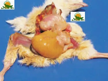
5. At a later stage of the infection, the yolk content is a cause of putrefactive necrotic processes in the peritoneal cavity. The abdomen is bloated. The entire abdominal wall is affected by a moist gangrene (maceration).

6. Salpingitis (inflammation of the oviduct). Salpingites due to E. coli infections could be also observed in growing birds. The oviduct is dilated, with thinned wall and filled with caseous exudate all along its length.


7. Salpingites are among the commonest causes for death in layer hens. E. coli penetrates from the cloaca via an ascendant route. Predisposing factors are the intense egg laying and the associated estrogen activity.


. 8. Salpingitis. In older cases, the caseous masses in the oviduct have a lamellar structure. E. coli organisms are usually found in excreta because of their presence in avian and mammalian intestine, the birds are constantly at risk of infection through contaminated water, dust, faeces and environment.


9. Salpingitis. Retained yolks among the caseous masses in the oviduct. In some cases, when the systemic resistance is lower, places, contaminated with E. coli, such as intestine, genital tract or nasal passages, could be latent sources of infection.


10. Compression and compactness of caseous necrotic masses after losing a part of their water content in the oviduct of a layer


11. Egg yolk peritonitis in a layer hen consequently to E. coli salpingitis. The chickens could be hatched with a latent infection, when E. coli is present in ovaries and the oviduct. In these instances, the infection could turn into an overt infection under the influence of some stress factors or lesions.

12. (inflammation of the ovary) consequently to a salpingitis due to ascendant E coli infection.

13. Cystic degeneration of ovarian follicles following an E. coli opphoritis.

14. Cellulitis (inflammation of the subcutaneous tissue that affects also the overlying skin). It predominates in broilers and is detected mainly in slaughterhouses. Macroscopically, the lesions are with a yellowish-brown colour.

15. Cellulitis. Affected areas are mostly in the region of the back and the thighs.

16. In some cases, the lesions are slightly prominating over the adjacent healthy skin.

17. Cullulitis. In the subcutaneous tissue, thick fibrinous plaques are often found out.

18. In some cases with adult birds, in the region of the head, subcutaneous masses of thick serofibrinous exudate resulting from a local E. coli infection could be detected.

19. Enterocolitis. Enterotoxigenic E. coli that produce toxins, cause the secretion and retention of fluids in some intestinal loops and especially in the caeca. Clinically, diarrhoea and de¬hydration are observed. The intestines are pale and distended, particularly the caeca that are overfilled with fluid containing many gas bubbles

20. . Neonatal E. coli septicaemia. Chickens in the first 24 - 48 h after hatching are affected. The death rate during the first ten days is higher and could reach 5 - 6%. The yolk sac is unabsorbed. The spleen is enlarged. Some days later, the typical serofibrinous polyserositis lesions, affecting the peritoneum, the pericardium, the air sacs and the liver capsule are manifested.

21. Acute E. coli septicaemia in layer hens. Clinically and morphologically, the acute E. coli septicaemia could resemble fowl cholera or fowl typhoid. It is encountered in both young and sexually mature birds. The stress in the beginning of egg-laying is considered as an important predisposing factor. The parenchymal organs are enlarged and hyperaemic. Sometimes, the liver has a greenish color and is mottled with multiple small necrotic foci. Also, pericarditis, peritonitis and petechial haemorrhages on serous coats are present.


22. E. coli septicaemia of a respiratory origin. In such cases, the respiratory mucosa damaged by infectious and non-infections agents (ND viruses including vaccinal strains, IB, TRT, mycoplasmae, high ammonia levels) is the entrance door of the E. coli infection. The lesions are principally observed in the respiratory tract (trachea, lungs and air sacs), but some adjacent serous coats (pericardium, peritoneum) are also affected and thus, the picture of a typical serofibrinous polyserositis is produced.

23. E. coli septicaemia secondary to enteritis. It is most commonly encountered in turkeys. The intestinal mucosa, damaged by the haemorrhagic enteritis virus (see Adenovirus infections), is the entrance door of E. coli infection. The most typical lesions are the marked enlargement, hyperaemia, haemorrhages and necrosis of the liver and the spleen.

24. Panophthalmitis (inflammation of all tissues of the eyeball). Generally, it develops secondary to E. coli septicaemia and is usually unilateral.

25. Arthritis, Osteomyelitis and Osteonecrosis (inflammation of joints, bone marrow and bone necrosis, respectively). The lesions are a common sequel to E. coli septicaemia. Clinically, lameness, prolonged lying down, dehydration and retarded growth rate are observed. The coxofemoral joints, the femur and tibiotarsal joints are most commonly affected. The bacteria colonize the physes of growing bones and provoke an inflammatory response that is further causing osteomyelitis. Pathoanatomically, fractures of the femoral head are usually discovered.

26. some cases of E. coli septicaemia, accumulation of exudate between the superficial and deep pectoral muscles is observed.

27. The lesions that develop in the articular spaces of thoracolumbar vertebrae result in spondylitis (spondylosis) and after that, in progressive paresis and paralysis.

28. Coligranuloma (Hjarre's disease). It is characterized by multiple granulomas in the intestinal tract, the mesentery and the liver, but not in spleen. The lesions are similar to these observed in tuberculosis.


29. Bursitis sternalis (inflammation of the sternal bursa). The bursa is enlarged in a various extent and filled with inflammatory exudate. The diagnosis of coli - infections is based on isolation and typization of pathogenic E. coli serotypes. Many other bacteria (salmonellae, pasteurellae, staphylococci etc.), viruses, chlamydiae and mycoplasmae should be excluded as possible aetiological agents. The prevention should aim at minimizing the probability of faecal contamination of eggs. This implies the maintenance of clean nests, discarding floor eggs and removal of eggs that are cracked or contaminated with faeces. Breeder eggs should be fumigated or disinfected in the farm prior to their transportation in the storage premise. The treatment is effective if initiated soon after testing the antibacterial sensitivity of isolates.

Key points of Poultry diseases

Disease
Etiology
Important points
Avian tuberculosis
Mycobacterium avium

Older birds usually affected ,Tubercles on different organs .Prominent keel bone ,acid fast staining

Avian spirochetosis
Borelia anserina
 High body temperature. Watery greenish diarrhea
Presence of Argas percicus, bluish comb ,splenomegaly 
Clostridial infections
Necrotic enteritis
Cl.Perfringens
 Reddish dark color diarrhea  that is some time confused with coccidiosis ,Turkish towel appearance
Ulcerative enteritis
Cl.Colinum
Ulcers on intestine ,watery diarrhea
Gangrenous dermatitis
Cl.Septicum 
Gases and serosanguinus fluid under skin, gangrene area under skin 
Botulism
Cl.Botulinum
Limber neck ( flaccid paralysis)
Salmonella infections
Pullorum disease
Salmonella Pullorum
Young age, non-motile, host specific, chalky white diarrhea ,mushy chicks ,hock joint abscess, cecal cores,10-15 % egg infected. Horizontal + vertical transmission.
Fowl typhoid
Salmonella Gallinarum
Host specific non motile, Sulphur color diarrhea ,grower and adult birds
Bronze coppery color liver, 80-90% egg infected Horizontal + vertical transmission.
Fowl paratyphoid


Salmonella typhimurium
Salmonella enteritidis
Motile, non host specific, watery diarrhea, young age, cecal casts, button type nodules on intestine Horizontal + vertical transmission.


Collobacilosis


E.coli


Motile, pan systemic disease, coli septicemia, coli granuloma, Hjjarre’s disease in turkeys
White fibrin layer on heart, liver, CCRD
Infectious Coryza
Hemophilus Paragalinarum
Disease of URT, foul smelling discharge from eyes and nostrils ,sticky and watery eyes. Roup (weeping birds)conjunctivitis ,sinusitis ,facial edema ,swollen head ,swollen comb and wattles
Fowl cholera


Pastreulla multocida
Adult birds, swollen wattle, edema of throat.
Watery greenish diarrhea ,hunched up position,discharge from mouth Corn meal liver

Mycoplasmosis (CRD)
Mycoplasma gallisepticum
URT+ LRT, airsaculitis ,cheesy exudates on air sacs ,foamy appearance of air sacs .Frey’s medium, PPLO broth  .fried egg colonies ,CCRD ,razor blade keel bone .Horizontal + vertical transmission .
Brooder pneumonia
Aspergillus fumigatus,
aspergillus flavis
Gasping ,miliary tubercle on lungs (pin head to pea size) usually brooding age
Candidiasis (thrush)
Candida albicane
Ulcer in mouth & esophagus ,crop .curd like appearance of crop ,thickened crop
Mycotoxicosis
Aflatoxins,
ochratoxin,
trichothesens,
etc.
Atrophy of immune organs, (bursa, thymus) pale swollen hemorrhagic liver. Hemorrhages on body. koilen layer easily peeled off .
Bloody thigh syndrome ,lazy leukocyte syndrome
Coccidiosis
E. acervulina
E. brunette    
.E. praecox
E. maxima E. mitis
E. mivati     
  E .necatrix
E. hagani      
 E.tenella

Host specific, site specific, Pin point hemorrhages on intestine (Frank hemorrhages), blood in intestine ceca   become engorged with blood. Bloody diarrhea.
Black head (Histomoniasis )


Histomonas meleagridis
Characterized by black head & circular necrotic liver lesions ( Bull eye lesions )

Internal parasite of poultry GIT
Cappilarea annulata, cappilarea contorta (cappilariasis ) Ascaradia galli (ascariasis) Davenia, Rellitena (tapeworm)Hetarekis gallinarum
Less efficient utilization of feed. Hemorrhagic enteritis, diarrhea, emaciation, loss of weight & egg production. Intestinal blockage in severe infections. Occasional finding of A. galli in hen’s egg due to migration of worm to oviduct via cloaca & subsequent inclusion in egg.

Internal parasite of poultry respiratory system
Syngamus trachea
Gap worm/
( Gap,syngamiasis)
/Forked worm/
Y-shaped worms
Diffuse or focal tracheitis, gasping, dyspnea & head shaking due to physical blockage of airways. Signs resemble laryngotracheitis


Rapid Mycotoxin Test / "ELISA Test"


8. Rapid Mycotoxin Test / "ELISA Test"

Principle :

Antibody coated column is used to trap the mycotoxin. This trapped toxin is then eluted using approximate solvent and quantified in fluorometer.

Equipments :

  • Immuno affinity column 
  • Affinity column stand with syringe 
  • Cuvette 
  • Calibrated Fluorometer 
  • Blender 
  • Fluted filter paper

Reagent:

  • Test developer 
  • Methonol : water (80 : 20 by volume) 
  • Mycotoxin wash buffer

Procedure:

  • 50 gms of sample + 5 gms of NaCl + 100 ml of methanol water (80 : 20)                                        Note : NaCl is not added in case of Ochra Test
  • Blend at high speed and filter
  • Pipette filtered extract into clean vessel Aflatoxin, Ochratoxin : 10 ml
  • Zearalenone : 1 ml
  • Dilute with purified water and mix                                                                                                                Aflatoxin, Ochratoxin : 40 ml                                                                                                Zearalenone : 49 ml
  • Filter
  • Remove top cap and attach the syringe (cut 1/8 inch bottom of column )
  • Pass filtered diluted extract at the rate of 1-2 drops/second                                                                             Aflatoxin : 2 ml Ochratoxin,                                                                                                 Zearalenone : 10ml
  • Pass water at the rate of 1-2 drops/second                                                                                                    Aflatoxin, Zearalenone : 5 ml                                                                                                Ochratoxin : First 10 ml Mycotoxin wash buffer,                                                                     Later 10 ml distilled water                                                                                                                                         Courtesy : VICAM
  • Elute toxin in glass cuvette                                                                                                                        Aflatoxin, Zearalenone : Pass 1 ml HPLC grade methanol                                                        Ochratoxin : Pass 1.5 ml Ochratoxin eluting soln.
  • Add 1 ml of developer to the cuvette and mix well
  • Read in calibrated fluorometer

Bacterial Diseases

Bacterial Diseases:-
Hemorrahgic septicemia(H S):
Also known as pasturelosis, stockyardis disease, shiping fever, barbon diseaseThis is No 1 bacterial disease in Pakistan. H.S is an acute ,septicemic bacterial disease of cattle and buffalo. It is characterised by high temp, severe depression, pneumonia and swelling at throat region/ neck.
Etiology:
            Pasturela Multocida is the causative agent of this disease. It has four types. Type A: It causes

  •                            Bovine pneumonic pasturelosis in cattle and buffalo.
  •               Mastits, pneumoing in sheep and gaot.
  •               Rhinits and pneumonia in Pig.
  •               Fowl cholera in poultry.

Type B: it causes Hemorrhagic septicemia in Asia.
Type D: causes rhinitis in pig
Type E: causes Hemorraghic septicemia.
Predisposing Factor:

  • Over work that cause fatigue,
  • Poor body condition,
  • Moonsoon rain,
  • Transportation stress
  • close confinement
Epidemiology:
            H. S is present in all tropical and subtropical areas. Mostly it occurs in Rushia,  Africa, Australia, India, Pakistan, Indionasia, Malayasia, Phelpine and Thiland.
 Buffalo is most susceptible than cattle.
Animal of all age are susceptible but more susceptible age is 6-24 months.
 Mortality and Morbidity rate vary from 50-100%.
Out breaks of this disease is often associated with wet humid wheather during rainy season.
Pathogensis:
            Pasturela Mulltocida normally commensel of upper respiratory tract and may invade tissue of immunosupressed animal. Exogenus transmission occurs through direct contact or through aerosol route.
2 factors are important in pathogensis .

  1. adhesion of bactera to mucosa (fimbrae).
  2. To avoid from phagocytosis (capsule).
             Severe septicemia may occur which is associated with capsular material of bactera. The effect of septicemia are more severe in respiratory tract, heart and gastrointestinal tarct.
Clinical Findings:
  Four type of clinical findings.

  1. Oedematous form
  2. Pulmonary form
  3.  Alimentary  form
  4. Septicemic form                                                                                                               
1. Edematous form:

  •  Swelling on neck/throat region.
  • High temperature upto 106-107 F.
  • Off feed
  • Severe depressed
  •  Perfuse salivation
  • Dyspnea
  • Some time death may occur in severe cases.
2.  Pulmonary form:

  • No swelling on neck
  • Temperature remain 104-105F
  • Open mouth breathing
  • Protrude and cyanotic tongue
  • Dyspnea which may lead to death.
3. Alimentary form:

  • Diarrhea
  • Loss of appetite
  • Increase thirst
  • Dehydration
4. Septicemic form:
  •  Increase temperature
  • This form is of very short duration, endotoxin produce and circulate in blood. Animal die in short period of time
Diagnosis:

  • History and clinical sign
  • Observe throat swelling
  • Open mouth breathing
  • Tongue cyanotic
  • Observe month( june, july and august.
Differential diagnosis:

  1. Black Quarter:

  •    clostridium infection, thigh muscle and shoulder muscle involve.
  •    Lameness
  •    Swelling of quarter
  •    This is soil born disease.
     2. Anthrax:
  • Bleeding from natural orifices
     3.Wooden tongue:
Treatment:
Use bacteriostate drugs instead of bacteriocidal in this disease.
Drug of choice is Sulphonamides calve 100ml large animal 200ml I/V
Or
Tribersin (sulphmetnzine 150mg/kg + chloramphenicol 10mg/kg) for 3-4 days.
Give fluid therapy.
Use antipyretic .
Use bronchodilators

Mastitis:-

It is an infectious but consider as managemental disease. Mastitis is characterized by inflammation of parenchymal cells of udder.
Inflammation of mamory gland is called as mamilitis. Complication of mamalitis is mastits.
Etiology:-
Staplococcus aureus is gram +ve bacteria and other bacteria like streptococcus agalacae, corynobacterium, E. coli, enterococci, mycoplasma bovis, klebshiela etc.
Mastitis is actually contagious and environmental disease.
Managemental problems:-
            Poor bedding, housing, hot, humid area favour pathogens present in wood dust, rice straw.
Mortality is negligible but morbidity is very high and more economic loss.
Subclinical mastitis has more economical losses because no signs so no treatment.
Physio-chemical changes in mastitic milk are taste, consistency. Only can be diagnosed by tests.
Clinical signs:-
Subclinical mastitis:-
Anthrax: 
It is also known as spleenic fever, carbuncles, wool sorter’s disease, hide porter’s disease and malignant pustular dermatitis.
It is per acute disease characterized by septicemia and sudden death accompanied by the exudation of tery blood from the natural orifices after death.
Etiology:
This disease is caused by bacteria bacillus anthracic. Bacillus anthracic is Gram +ve , spore forming, non-motile capsulated, aerobic bacteria. Spore are very resistant to chemical disinfectant. These spore may survive in the soil for years and for ten years in the culture. The organism produces medusa head appearance on nutrient agar. The spores remain resistant to 100oC for five minutes.
Epidemiology:
Anthrax is world wide in distribution but incidences are more common in hot-humid climate. Infection remains in the soil and suitable climate supports the occurrence. The morbidity and mortality rate is very high.
Pathogenesis:
After ingestion of the spores or entry through abrasions in the epithelium or through scratches caused by fibrous food materials. The bacteria are moved to the local lymph nodes by motile phagocytes. After proliferation in this site the bacilli pass via the lymphatic vessels in to the blood stream and septicemia takes place followed by massive invasion of all body tissues. During multiplication it releases lethal toxins that cause edema, tissue damage, and haemorraghes. Death occur due to shock, renal failiure and terminal anoxia mediated by CNS.
Clinical findings:
*      Incubation Period: 2-10 days
Four types:

  1.  Per acute
  2.  Acute
  3.  Sub-acute
  4. Chronic
1. Per-acute:

  •       Death occurs without showing any signs.
2. Acute:
  • Temp 104 oF
  •  Increase heart beat
  • Rapid and deep respiration
  • Mucosae congested and hemorrhagic
  • Diarrhea and dysentery
3. Sub-acute and Chronic forms:
  • Edema
  • Loss of appetite
  • Blood in milk for weeks
Postmortem findings:

  • Carcass should not be opened if confirmed
  • Terry blood from natural orifices
  • Increase spleen size
Diagnosis:

  • On the basis of signs
  • History
  • Ascoli’s test for anthrax
Differential diagnosis:-
Black Quarter: 

  • there will be frothy bleeding from nostrils. But in anthrax there is terry blood from natural orifices.
Treatment:
Pencilline 10000 IU/kg I/M twice daily is drug of choice
Oxytetracycline 8-10 mg/day

Black Quarters:-
            It is also known as black leg, quarter ill and symptomatic anthrax.It is manifested by severe toxemia and high mortality.

Etiology:-
            It is caused by clostridium shovei which is gram +ve, rod shape, anaerobic and spore forming bacteria. The spores of this bacterium are highly resistant to heat. Spores can tolerate the temperature up to 120oc or may live for many years in the soil. This bacterium have four type of exotoxins  α, β, γ and epsilon toxins.
Epidemiology:-
            Bollinger was scientist who first who identify this disease. Black quarter is an enzootic disease of tropical countries of world. This disease occurs after heavy rainfall and flood. Animal of 6-24 months are more susceptible for this disease. While cattle is more susceptible than other animals. Animal having good body condition are more susceptible to this disease.
Transmission:-
            This disease comes under the heading of soil born infections. It is spread by infected carcass. It may also be transmit due to contaminated feed, water, wound or trauma in large animals. In small animal it may be transmitted during shearing, docking, castrating or kidding/lambing.
Pathogenesis:-
            Spore may enter through injured mucosa of alimentary tract then it moves to the blood where it produces toxins and causes septicemia. Then it is engulfed by macrophages and reaches to muscles and causes hemorrhages due to injured capillaries. Toxins produce systemic reaction and local lesions and local lesions. Aspirate toxemia cause death in animals. In immune animals there is permanent damage of affected part.
Clinical signs:-
1.      Incubation period: 2-6 days
2.      Increase temperature 106-107 oF
3.      Swelling of affected part………. Swelling is hot and painful with passage of time swelling will cold and painless.
4.      Bubble formation by toxins.
5.      Necrosis
6.      Animal will feel restlessness
7.      Dull, depress
8.      Loss in appetite
9.      Abdominal pain and increase thirst
10.   Skin become dry and rough
11.  Death occur within 3-4 days
12.  In severe cases there may be lemeness and rupter of skin, discoloration of skin and fowl smelling.
Diagnosis:
            O n the basis of signs and symptoms
            FAT ( florescent antigen test)

Differential diagnosis:-
            Antrax:- at the death there will be bleeding in natural orifices. But in Black Quarter there will be frothy bleeding from nostrils.
Treatment:-
            Pencilline is drug of choice 20 I.U twice daily
            NASAIDs or steroids 1ml/10kg
            Antihistaminic drugs (Avil)
            Fluid therapy
            Multivitamins (never use ca in high fever because it leads to tachycardia.)
Controls:-
            Vaccination in April
            Deep burial of carcass 6-7 feet.
            Avoid animal grazing on contaminated pasture.
            Never open the carcass


Tetanas:
       It is also known as lock jaw, saw horse disease.

Etiology:
       Tetanus is caused by clostridium tetani. It is a G +ve, motile anaerobic bacteria. It is normal habitat in carnivors. It has three strains H, A and O. and produces three types of toxins.

  1. Tetanospasmin (neurotoxin)
  2. Tetanolysin (haemolysin)
  3. Fibrolysin
Epidemiology:
           Tetanus occurs in all species of animals. All age of animals such as horses, mules, sheep, goats and cattle are susceptible but equines are more susceptible among all. Sometimes outbreaks are observed in young animals. Morbidity rate is high and mortality rate is 80%.
Pathogenesis:
             The bacteria enter through deep punctured wounds in case of retain placenta, porlapse, shearing, docking and vaccination. When there is low Oin these tissues, this will help the spores of clostridium to germinate, grow and produce toxin. Tetanospasmin or neurotoxin is released by cellular autolysis. It is absorbed and disseminated in to bold to produce neurotoxic effect through absorption by peripheral nerve endings. It affacts centrally in the spinal cord and brain and peripherally on motor nerves to produce spastic paralysis. Flaccid paralysis also occurs due to local invasion of the nerves. Death occurs due to apnea as a result of fixation of respiratory muscles.
Clinical findings:
  • Incubation period few days to few weeks.
  • Suddenly progressive stiffness.
  •  Prolapsed of  3rd eye lid with the passage of time.
  •  Erection of ears
  • Saw-horse posture.
  • Animal standed with extended feet.
  • Death occur due to asphyxia
Diagnosis:
  •  on the basis of history
  •  clinical signs
  • stiff legs and locked jaw.
Differential diagnosis:
Milk fever: 
             In milk fever there quick response of ca therapy but in tetanus there is no such effect of calcium.
Strychnine poisoning: 
              History of accident feeding, absence of wounds,
Treatment:
Pencilline 10000IU/kg twice
 Chlorpromazine 0.2mg/kg as muscle relexant
 Mgso4
Homeopathic treatment:
Camphora-10 drops every 20 minutes. After camphora administer belladonna every hour.

Lpetospirosis:
Etiology:
           Leptospirosis is caused by leptospira, which is more than 212 serotypes. In old books it is serotype in new one it is serovars. The pathogenic specie is L. enterogen. It affects all ruminents, canine and feline family as well as in human beings. This disease is of two types
  1. Host adapted
  2.  Non host adopted
Non host adopted:
L. pomona is specific for dogs if it infect in cattle that infection will be non host adapted infection. The cattle will be incidental or accidental host.
Host adapted:
L. hardjo is specific for cattle. If cattle get infected from this then it will be host adapted infection. Non host adapted form of infection is more severe than host adapted infection.
Transmission:
          This is more fragile bacteria; it grows in moist, marshy and rainy areas. It can servive upto 6 month. It destroyes with in 30 min if the area is dry. PH from 6-8 is gud growth for its growth. It cannot servive in less than 10 C and more than 40 C. it shed through urine, semen, aborted fetus. Sheding occur after 6 month of recovery from disease.
Pathogensis:
             It enters through skin, ingestion and direct contact and multiplies in epithelial cells and extra cellular fluid. Then it enter into blood and produces hemolysin and cause hemolysis. its predilection site is liver and kidney. In kidney it causes glumerulonephritis. It also causes abortion in 2nd trimester of pregnancy.
This is No# 1 occupational hazzard in newzeland. 34% veterinarians are infected with Leptospira. No vaccine available for human.
Clinical Findings:
Three types
  1. Acute
  2. Subacute
  3. Chronic
Acute:
  •  I.P= 4-7 days
  • Temperature upto 107 F
  •  Milk production drop
  •  Anorexia
  • Anemia
  • White spotted kidney
  • Dyspnea
  •  Abortion in 1st week
Subacute:
  •  Less severe
  • Abortion in 3-4 weeks
  • Increase temperature upto 105 F
Chronic:
  • Abortion and other signs
Occult:
  • Only abortion
  • Increase antibody titer
 L. pomono causes acute and subacute form it secretes hemolysin and causes homolysis.
L. hadjo cause chronic form it cannot secrete hemolysin but causes abortion.
Differential diagnosis:
1. Bacillary Hemoglubinuria:
No abortion and is caused by clostridium hemoluticam, clostridium novyae. No milk drop syndrome spountaneosly.
2. Bebasiosis:
Ticks are present.
3. PPH:
Due to the deficiency of Phosphorus. No temperature.
Diagnosis:
Culture tests.
Treatment:
Streptomycin, ox tetracycline are drug of choice.
Conrol:
         Vaccine is available for dog, buffalo and cattle. Pentavalent vaccine for cattle and buffalo. L. Pomona, L. hardjo, icterohemorrhagica, kirschneria and grippotyphosa.
Tetravalent vaccine is for dog.

Bacillary Hemoglubinuria:
     It is caused by clostridium hemolyticum, clostridium  navyae type D. spore forming anaerobic soil born rod shaped G +ve bacteria. It is normally present in the soil and liver. All clostridail pathogens live in soil as well as in liver. They can survive up to 1 year outside the body of animal. This was observed on the bone of dead animal which was died one year ago.
Clostridium Navyae type D produce two types of toxin.
  1. Necrotoxins
  2. Β- hemolytica
Type A, B; C and D are toxin producer. All are pathogenic except type C which is non pathogenic. Type B cause black disease, necrotic hepatitis in sheep and goat.
Necrotoxins:
It causes necrosis.
Β-hemolytica:
it causes rupture of blood vessels, breakdown of RBCs.
This disease is prevalent in autumn and summer.
Transmission:
Transfer through direct ingestion from there it goes to epithelial cells of liver (predilection site). If damage of parenchyma it causes anerobic condition and start its activity and cause localize necrosis.
Clinical Findings:
  •   Ip 1-7 days
  •  Sudden death without signs.
  • Sudden increase temperature up to 106-107F.
  • Sudden decrease in milk production
  • Animal feel abdominal pain due to liver damage
  • Edema of braskit
  •  Dark brown color feces
  •  Hypoxia and severe anemia
  •  Death
  • Abortion in later stages.
Treatment:
  •  G +ve medication.
  • Pencillin drug of choice for all clostridium pathogens
  •  Amoxicilline
Control:
  • Vaccination 4-6 week before prevalence
  • In calf and newborn vaccinate at six month and then annually
  • Twice in year if the disease is more prevalent
  • Affected carcass should be properly disposed off.
Actionmycosis (Lumpy Jaw):
Actinomycosis is sporadic disease therefore rarely animal transmission